
Bác sĩ giải đáp: Khám thai lần đầu có cần siêu âm đầu dò không?
Khám thai lần đầu có cần siêu âm đầu dò không? Bác sĩ giải đáp khi nào chỉ định và siêu âm đầu dò có an toàn không, khác gì siêu âm thành bụng. Xem ngay!
Many expectant mothers feel anxious before their first prenatal visit, especially when the doctor suggests a transvaginal ultrasound. The most common worries are: does the first prenatal visit really need a transvaginal ultrasound, is it mandatory, does it affect the baby, and can it be replaced by an abdominal scan?
Not every case calls for a transvaginal ultrasound. The doctor chooses the method based on gestational age, the mother's symptoms, and the goal of the visit. When the pregnancy is still very early, a scan through the vagina usually gives a clearer image, helping to confirm the exact location of the pregnancy and its early development.
This article was medically reviewed by Dr. Pham Thi Huyen — Department of Diagnostic Imaging, NeoMedic Medical Center — who will help you find the answers to these questions.
Does the First Prenatal Visit Need a Transvaginal Ultrasound?
Not every first prenatal visit requires a transvaginal ultrasound. Choosing between a vaginal scan and an abdominal scan depends on gestational age, hCG levels, clinical symptoms, and the obstetric assessment goal.
For a pregnancy that is only a few days past a missed period, or around 4–6 weeks, a transvaginal ultrasound is usually preferred. Because the probe sits close to the uterus, it produces more detailed images than an abdominal scan. This lets the doctor identify the gestational sac, estimate gestational age, and detect early abnormalities such as an ectopic pregnancy or a non-developing pregnancy.
When the pregnancy is further along — especially after 7–8 weeks, once the gestational sac and fetal heartbeat are clearly visible through the abdominal wall — an abdominal scan is usually enough for assessment, with no need to switch to a transvaginal scan.
For example, a woman whose period is 10 days late tests positive on a home test and comes in for her first visit. An abdominal scan does not yet show the gestational sac because the pregnancy is too small. On the transvaginal scan, the doctor can see the gestational sac correctly positioned inside the uterine cavity and schedules a follow-up in a few days to check for the fetal heartbeat. This is a fairly common situation in obstetric practice.
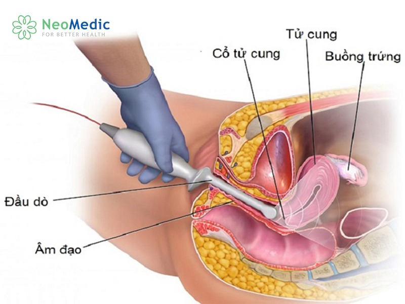
Not everyone needs a transvaginal ultrasound at their first prenatal visit
Quick summary:
- Not every case needs one.
- The earlier the pregnancy, the more likely a transvaginal ultrasound is needed.
- The doctor chooses the appropriate method after the examination.
- The goal is to assess the pregnancy accurately and keep both mother and baby safe.
According to the American College of Obstetricians and Gynecologists (ACOG) and the International Society of Ultrasound in Obstetrics and Gynecology (ISUOG), transvaginal ultrasound is highly valuable in the first trimester for confirming the location of the pregnancy, gestational age, and the pregnancy's viability, at a stage when an abdominal scan does not yet provide enough information.
When Doctors Order a Transvaginal Ultrasound at the First Prenatal Visit
Being pregnant does not automatically mean you need a transvaginal ultrasound. The doctor weighs the diagnostic benefit against each woman's condition before choosing the method. Most indications fall into the three groups below.
The Pregnancy Is Very Early and the Abdominal Scan Isn't Clear
In early pregnancy, especially before 6 weeks, the gestational sac is still very small. Ultrasound waves passing through the abdominal wall may not capture a clear image, particularly in women with a thick abdominal wall or an insufficiently full bladder.
A transvaginal ultrasound brings the probe closer to the uterus, making it easier to detect the gestational sac and yolk sac and to estimate gestational age more accurately. This method is also used when the pregnancy's development needs to be monitored during the first weeks.
Suspected Ectopic Pregnancy
An ectopic pregnancy occurs when the embryo implants outside the uterine cavity, most often in the fallopian tube. Detecting it early helps the doctor choose the right treatment approach and reduce the risk of complications.
A missed period accompanied by one-sided abdominal pain, abnormal vaginal bleeding, or an hCG level that rises out of proportion to the gestational age are situations in which a transvaginal ultrasound is often ordered. This method helps determine whether there is a gestational sac inside the uterine cavity, while also examining the adnexa for signs suggesting an ectopic pregnancy.
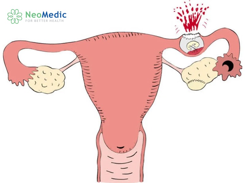
Suspected ectopic pregnancy
Abdominal Pain or Bleeding in the First Trimester
Dull abdominal pain, cramping, or vaginal bleeding in early pregnancy all need to be assessed early. The cause may be a normal feature of pregnancy, but it may also be related to a threatened miscarriage, an ectopic pregnancy, or a non-developing pregnancy.
These cases are usually given priority for a transvaginal scan because it provides more detailed images than an abdominal scan. The doctor checks the location of the gestational sac, the appearance of the yolk sac, the fetal heartbeat, and the condition of the uterine cavity to find the cause.
For example, a woman at around 5 weeks has brownish bleeding with mild abdominal pain. The transvaginal ultrasound shows the gestational sac inside the uterine cavity but no embryo yet, because the pregnancy is still early. The doctor schedules a follow-up in 7–10 days to assess development, rather than concluding an abnormality at the very first visit.
What Problems Can a Transvaginal Ultrasound Detect in Early Pregnancy
A transvaginal ultrasound is most valuable in the early weeks, when an abdominal scan is still limited. Beyond confirming the pregnancy, this method helps the doctor detect several important issues early so they can be monitored appropriately.
Confirming the Gestational Sac and the Location of the Pregnancy
One of the first goals of a prenatal visit is to determine whether the gestational sac is inside the uterine cavity.
If the gestational sac is already inside the uterus, the risk of an ectopic pregnancy drops considerably, although some special cases still require further monitoring. Conversely, when the hCG level has reached a threshold but no gestational sac is seen inside the uterine cavity, the doctor will consider whether the pregnancy is simply too early or whether an ectopic pregnancy is suspected.
Assessing the Yolk Sac and the Embryo
After confirming the gestational sac, the doctor looks next at the yolk sac and the embryo. These structures reflect the early development of the pregnancy.
The presence, size, and shape of the yolk sac help assess the pregnancy's potential to develop. If no embryo is seen while the pregnancy is still early, the mother is usually scheduled for a repeat scan in a few days, to avoid a premature conclusion.
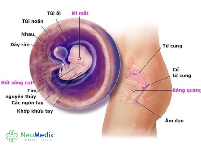
Assessing the yolk sac and the embryo
Checking the Fetal Heartbeat and the Pregnancy's Development
At the appropriate stage, a transvaginal ultrasound can detect fetal heart activity before an abdominal scan can.
Detecting a fetal heartbeat both confirms that the pregnancy is developing and helps the doctor assess the pregnancy's viability at the time of the exam. However, not seeing a heartbeat when the pregnancy is still very small does not confirm an abnormality. The doctor combines gestational age, the ultrasound findings, and the hCG level to plan follow-up.
Detecting an Ectopic Pregnancy Early
This is an important advantage of the transvaginal scan.
While examining the uterine cavity, the two adnexa, and the abdominal cavity, the doctor may notice signs suggesting an ectopic pregnancy: no gestational sac inside the uterus, an abnormal mass in the fallopian tube, or fluid in the abdominal cavity. An early diagnosis helps in choosing the right treatment plan and limiting complications.
How Does a Transvaginal Ultrasound Differ From an Abdominal Ultrasound?
Both use ultrasound waves, not X-rays, so neither exposes the mother or the baby to radiation. The main differences lie in where the probe is placed, the ability to visualize, and when each is used.
| Criterion | Transvaginal ultrasound | Abdominal ultrasound |
|---|---|---|
| Suitable timing | Very early pregnancy, first trimester | When the pregnancy is larger or already clearly visible through the abdominal wall |
| Image clarity | Clearer in early pregnancy | Depends on gestational age and the abdominal wall |
| Preparation | Usually no need to hold urine | May need a full bladder in very early pregnancy |
| Ability to detect the gestational sac early | Good | More limited when the pregnancy is still small |
| Level of discomfort | May feel slightly awkward during the procedure | Causes little discomfort |
Which method is chosen does not depend on the mother's preference but on the diagnostic goal. In many cases, the doctor starts with an abdominal scan; if the image is not clear enough, only then is a transvaginal scan added to provide more information.
According to ISUOG and ACOG, a transvaginal scan is recommended when early pregnancy needs to be assessed or when an abdominal scan does not provide enough imaging to reach a conclusion.
Does a Transvaginal Ultrasound Hurt? Is It Safe for Mother and Baby?
This is the most common worry at the first prenatal visit. Many people fear that inserting the probe into the vagina will touch the baby or increase the risk of miscarriage. These worries have no basis when the ultrasound is performed on the correct indication and with the correct technique.
Transvaginal ultrasound has been used widely in obstetrics for many years. The probe is small, covered with a sterile protective sheath, and inserted gently into the vagina. It stays only in the vagina and does not enter the uterine cavity, so it does not come into direct contact with the gestational sac or the baby. The method uses high-frequency sound waves to create images, not X-rays or ionizing radiation.
According to the American College of Obstetricians and Gynecologists (ACOG), the International Society of Ultrasound in Obstetrics and Gynecology (ISUOG), and NICE (UK), there is to date no high-quality scientific evidence that a correctly indicated diagnostic ultrasound, including a transvaginal scan, causes birth defects, increases the risk of miscarriage, or affects the baby's development.
| Tiêu chí | Siêu âm đầu dò | Siêu âm thành bụng |
| Thời điểm phù hợp | Thai rất sớm, ba tháng đầu | Khi thai lớn hơn hoặc đã quan sát rõ qua thành bụng |
| Độ rõ hình ảnh | Rõ hơn ở giai đoạn đầu thai kỳ | Phụ thuộc tuổi thai và thành bụng |
| Chuẩn bị | Thường không cần nhịn tiểu | Có thể cần bàng quang căng ở thai rất sớm |
| Khả năng phát hiện túi thai sớm | Tốt | Hạn chế hơn khi thai còn nhỏ |
| Mức độ khó chịu | Có thể hơi vướng trong lúc thực hiện | Ít gây khó chịu |
Dr. Pham Thi Huyen shares: "At 125 Thai Thinh Clinic, we had a woman 5 weeks pregnant who came in for mild abdominal pain. The doctor ordered a transvaginal ultrasound to rule out an ectopic pregnancy. After the scan, we confirmed the pregnancy was located inside the uterine cavity and advised her to continue routine prenatal monitoring. The transvaginal ultrasound caused no complications and had no effect on the mother's health or the baby's development."

A transvaginal ultrasound does not cause pain.
Does a Transvaginal Ultrasound Hurt?
Most pregnant women feel only slight fullness or mild discomfort in the first few minutes as the probe is inserted. The sensation is similar to a gynecological exam and eases as soon as the procedure ends.
The probe has a small diameter, is covered with a protective sheath, and is coated with lubricating gel before the procedure; the doctor works gently to keep discomfort to a minimum. Relaxing your body, breathing steadily, and following the instructions also help the process go faster and more smoothly.
Some women with a history of vaginitis, pelvic spasms, or a lot of anxiety may feel more discomfort. In that case, it's best to tell the doctor so the technique can be adjusted appropriately.
Can a Transvaginal Ultrasound Cause a Miscarriage?
No. According to current obstetric recommendations, there is no evidence that a transvaginal scan increases the risk of miscarriage in a normal pregnancy. Most first-trimester miscarriages are related to chromosomal abnormalities of the embryo or other medical causes, not to a transvaginal ultrasound.
After the scan, some women may have a little discharge or a few drops of light-pink blood. This is usually because the cervix is more sensitive during pregnancy, especially in women whose cervix bleeds easily or who have a cervical polyp. The amount of blood is very small and stops on its own after a short time.
If there is heavy bright-red bleeding, worsening abdominal pain, dizziness, or other unusual signs, the mother should return to a medical facility for a check. The cause is usually related to the state of the pregnancy rather than to the transvaginal ultrasound.
Can a Transvaginal Ultrasound Cause an Infection?
The risk of infection is very low when the ultrasound is performed at a facility that ensures infection-control procedures.
Every probe is disinfected according to regulations and used with a single-use protective sheath. The ultrasound gel is also a specialized type intended for transvaginal examinations.
When Should a Transvaginal Ultrasound Be Reconsidered or Avoided?
Although it is safe, a transvaginal ultrasound is not always the preferred choice. The doctor will assess the specific situation before ordering it. Some situations that call for reconsideration include:
- The pregnancy is already large enough for an abdominal scan to give a clear image.
- The woman declines the procedure after being fully counseled.
- Significant pain or vaginal injury makes inserting the probe difficult.
- Certain special situations that the doctor will assess individually to choose the appropriate technique.
In these cases, if the abdominal ultrasound image is clear enough, the doctor may prefer an abdominal scan over a transvaginal one.

A transvaginal ultrasound should be avoided once the pregnancy is large enough to see on an abdominal scan
What Can an Ultrasound Show Week by Week?
The ultrasound image depends heavily on gestational age. Scanning too early and not yet seeing all the structures of the pregnancy does not mean there is an abnormality.
| Estimated gestational age | Image usually seen |
|---|---|
| 4–5 weeks | The gestational sac may start to be seen inside the uterine cavity |
| 5–6 weeks | The yolk sac is usually seen; the embryo may appear |
| 6–7 weeks | In most cases, the fetal heartbeat is already visible |
| 7–8 weeks | The embryo develops more clearly; gestational age and heartbeat are assessed more accurately |
The milestones above are for reference only. The point at which each structure becomes visible can vary depending on the timing of ovulation, the menstrual cycle, and how each pregnancy develops.
For example, a woman with a 35-day cycle usually ovulates later than someone with a 28-day cycle. If gestational age is calculated from the last menstrual period, the ultrasound result may be a few days to a week smaller than expected while still being completely normal.
If no embryo or fetal heartbeat is seen at the first visit, the doctor usually schedules a repeat scan in 7–10 days for a more accurate assessment, rather than concluding a non-developing pregnancy right at the first visit. This is consistent with the recommendations of ACOG, NICE, and ISUOG for assessing early pregnancy.
Frequently Asked Questions About Transvaginal Ultrasound
Is a transvaginal ultrasound mandatory at the first prenatal visit?
No. The doctor will choose the ultrasound method that fits the gestational age and the purpose of the exam. If the pregnancy is already large enough to be seen clearly through the abdominal wall, a transvaginal ultrasound is usually not needed.
Does a transvaginal ultrasound affect the baby?
No. According to ACOG and NICE, a transvaginal scan is a safe method during pregnancy when performed on the correct indication and with the correct technique.
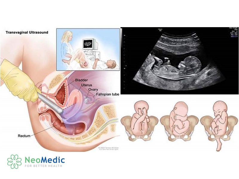
A transvaginal ultrasound does not affect the baby when performed with the correct technique
Does a transvaginal ultrasound hurt?
Most pregnant women feel only slight discomfort in the first few minutes. The procedure usually does not cause much pain and finishes quickly.
At how many weeks can the gestational sac be seen?
Typically, the gestational sac can be seen on a transvaginal scan from about 4.5–5 weeks of pregnancy, depending on the timing of conception and the hCG level.
At how many weeks can the fetal heartbeat be seen?
The fetal heartbeat is usually seen from about 5.5–6 weeks on a transvaginal ultrasound. In some cases it appears later if ovulation or conception occurred later.
If an abdominal scan doesn't show the pregnancy, is that an abnormality?
Not necessarily. If the gestational age is still small, an abdominal scan may not yet show the gestational sac. The doctor will usually order a transvaginal scan or schedule a repeat check in a few days.
If the fetal heartbeat isn't seen yet, is that a cause for concern?
Not always. If the pregnancy is still early or the menstrual cycle is irregular, the doctor will usually schedule a repeat scan in about 7–10 days, combined with monitoring the hCG level, before reaching a conclusion.
Does the first prenatal visit need a transvaginal ultrasound? The answer is that not every case does, but it is a very valuable method in the first weeks of pregnancy. When the pregnancy is still small, when an ectopic pregnancy is suspected, or when there is abdominal pain or vaginal bleeding, a transvaginal scan helps determine the location of the pregnancy and assess the gestational sac, yolk sac, fetal heartbeat, and the pregnancy's viability more accurately than an abdominal scan.
What matters is not whether you choose a transvaginal or an abdominal ultrasound, but being examined at the right time and on the right indication. If you have just had a positive pregnancy test or have unusual signs in early pregnancy, come to NeoMedic Medical Center to be examined and assessed by a specialist and to have the appropriate ultrasound method chosen for you.
Appointment booking: Book an appointment at NeoMedic Medical Center for a prenatal check and specialist advice on the right ultrasound method for you.
References:
- World Health Organization (WHO). WHO recommendations on antenatal care for a positive pregnancy experience.
- American College of Obstetricians and Gynecologists (ACOG). Ultrasound in Pregnancy.
- American Institute of Ultrasound in Medicine (AIUM). Practice Parameter for the Performance of Obstetric Ultrasound Examinations.
- ISUOG Practice Guidelines. Performance of first-trimester ultrasound scan.


